What is a CT Scanner? Are you sure this isn’t a Time Machine?
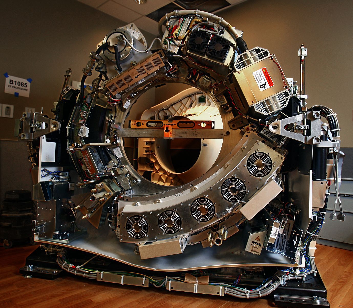
Well, CT stands for Computed Tomography. The machine is basically a traditional X-ray machine that spins around the patient so that it can acquire many different images of the body. A computer algorithm converts the images into layered scans to allow radiologists to sift through them looking for tumors, internal bleeds, pneumonia, and a range of other conditions. The final images look something like this.
CT scans are one of the most frequently ordered diagnostic tests in emergency departments in the United States (many say they are ordered way too much), so it’s important to know a little bit about how they work… And unfortunately, no, it is not a time machine.
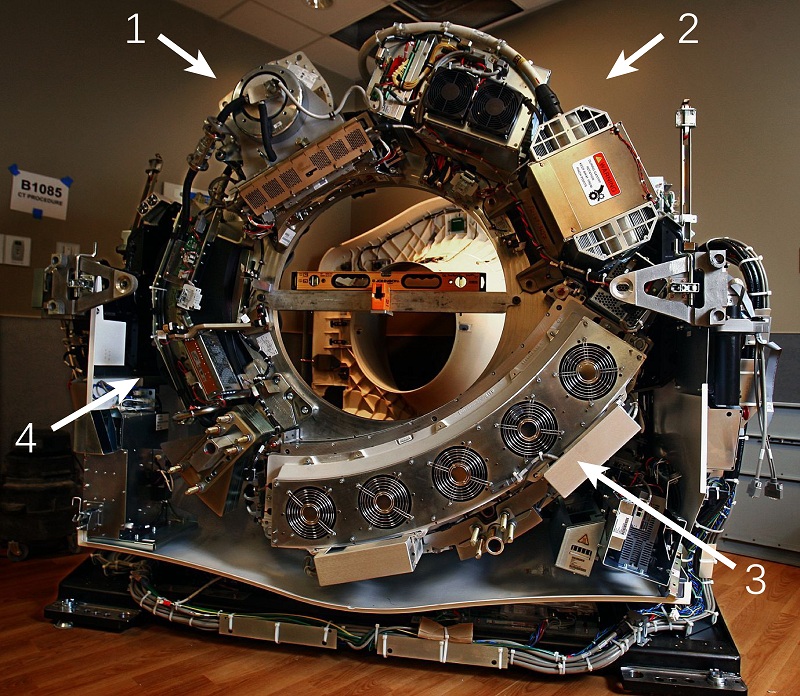
Here is a labeled image to give you a bit more detail into how it works:
4: Fluid pump and radiator for cooling the X-ray tube
All of these components make 2 to 3 complete turns per second around the patient.
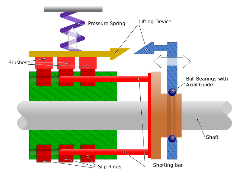
So how does the CT Scanner stay electrically powered while spinning?
To keep the machine charged without tangling the cords, CT scanners rely on the technology of the Slip Ring:
A Slip Ring is basically an electromechanical device that allows the transmission of power and electrical signals from a stationary to a rotating structure, in this case, from the base to the rotating scanner. One difference between the image below and the slip rings of CT Scanners is that there is a pool of liquid metal molecularly bonded to the contacts instead of the sliding brush. This decreases friction even more to allow constant rotation of the scanner.
Hopefully you found this interesting and at least somewhat easy to understand. For further reading, head here.
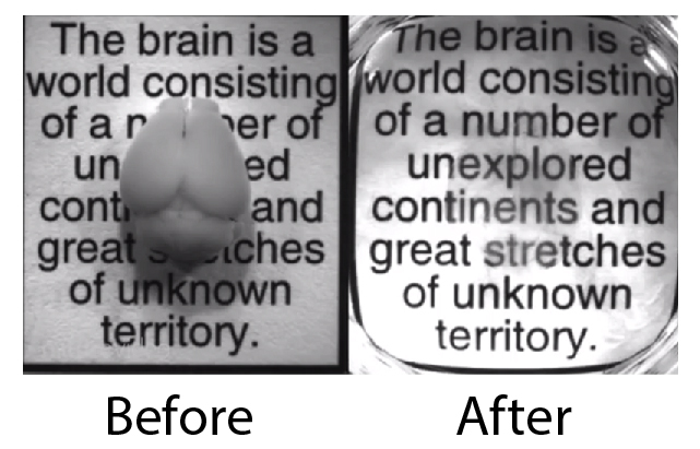
The world’s very first See-Through Brain has been developed by a team at Stanford University led by Karl Deisseroth (M.D., Ph.D.). Deisseroth is well-known for his critical role in the development of Optogenetics, a tool used to control individual neurons with light. Optogenetics is normally limited to surface neurons because the light has trouble reaching deeper areas, but the see-through brain may greatly enhance its efficacy.
The new method (termed CLARITY) involves removing the fat that provides structure but also blocks light. The brain is soaked in a chemical that forms a nanoporous hydrogel-hybridized mesh in the brain. This mesh can then support all the tissue so the fat can be washed away, resulting in the incredible see-through brain.
Unfortunately, the new technique can’t be used in living animals, but it still represents a huge advancement for neuroanatomists. No longer will there be much need to cut the brain into tiny slices (an extremely time-consuming process) to observe connectivity.
The announcement comes just a week after President Barack Obama announced a $100 million BRAIN initiative, and this new step forward surely offers a taste of the sort of technological breakthroughs the initiative hopes to achieve.
And all the Leaders in Neuroscience seem to be weighing in on this one:
“I can’t make any official statement, but I can say that this is exactly the type of technology one would hope to develop for the [BRAIN] project” – Dr. Michelle Freund, a program manager with the National Institutes of Mental Health
“If the entire mouse brain is transparent, that makes a very large fraction of neuroscience research much easier” – Dr. R. Clay Reid of the Allen Institute for Brain Science in Seattle.
This technique “is a giant step forward from having to slice the mouse brain into 1,000 pieces and looking at them each individually, then trying to reconstruct the relationships of all those slices” – Dr. Cori Bargmann of Rockefeller University, a co-leader of Obama’s brain initiative.
“It’s exactly the technique everyone’s been waiting for”- Dr. Terry Sejnowski of the Salk Institute.
Karl Deisseroth, mastermind of the CLARITY technique
It is certainly an exciting time to be a Neuroscientist.
Before reading this post, watch the 2 videos below:
If you’re like 50% of the people who watch the first video, you did not notice a gorilla pounding his chest in the middle of the scene. And if you are already familiar with that one, the second video can trick you in a new way. The basic idea of selective attention is that individuals have a tendency to process information from only one part of the environment with the exclusion of other parts. This can be extremely important in every day life. Consider for instance, you are driving through an intersection, and you are only looking for other cars that might hit you. buy sildenafil citrate tadalafil vardenafil online https://blackmenheal.org/wp-content/languages/en/sildenafil-citrate-tadalafil-vardenafil.html no prescription
While focusing on the cars, you may miss seeing a kid crossing on his bicycle.
And even more striking evidence for the importance of selective attention has recently come out of the Wolfe Lab at Harvard, it was demonstrated that radiologists may also suffer from this phenomenon at some level. The radiologists were given the image below…
and were asked to search for cancerous nodules in the image. Surprisingly, 83% of the professionally trained doctors didn’t notice a size-able gorilla shaking its arm at them.
This effect worked because cancerous nodules will show up as white circles on the image, so they are “inattentionally blind” to the black gorilla — the same reason you may have missed the gorilla in the video. buy strong pack online https://blackmenheal.org/wp-content/languages/en/strong-pack.html no prescription
Here’s how one commenter broke it down:
“I’m a radiologist. Air on xray/CT is black. The gorilla in this CT image is black. Black things in the lungs usually have no clinical significance. buy cipro online https://blackmenheal.org/wp-content/languages/en/cipro.html no prescription
Cancer is white. Pneumonia is white. Acute disease (other than a collapsed lung) is white. A collapsed lung is not in this location. While the “fact” that all the radiologists missed the gorilla may be shocking to lay people, the reality is that, given appearance/location/etc in this “experiment”, it just doesn’t matter.”
Gaining a better understanding of how our brain processes information can hopefully lead to safety nets that prevent mistakes.
Artistic rendering of a single pyramidal neuron of layer 5A of rat primary somatosensory cortex. These pyramidal neurons are involved in encoding whisker movement when the rat is actively exploring its environment. The part of the neuron receiving information from neighbouring neurons is shown in red; the part of the neuron sending information downstream is shown in blue. The green cylinders illustrate anatomical landmarks of the primary somatosensory cortex. buy levitra free viagra online https://apwh.org/wp-content/languages/en/levitra-free-viagra.html no prescription
The individual neurons are the basic units of the nervous system and through single cell reconstructions of these pieces of the puzzle; we aim to disentangle the intricate cortical microcircuit.
This may be my favorite.
J. Winnubst: Sleeper cell
When we are born our brains are already fully formed and will, to a large extent, determine our
feelings, personality and desires. In order to achieve this highly complex functionality, immature
neurons must venture out during their development and find the right cell partners to form synaptic
connections with. This process is aided by spontaneous network activity in the brain that tests and
refines the made connections. Some will stabilize while others are destined to be broken up. Shown
here is one of the ways spontaneous activity shapes and organizes connectivity: Synaptic inputs that
are close together on a neurons dendrite are more likely to carry similar information and are more
often co-active. The image illustrates how, even before the brain has become fully functional, a single
immature neuron is already tasked with finding order amongst the internal chaos of the mind.
The center of the image shows a labeled stretch of dendrite on which recorded calcium transients,
belonging to 2 co-activate synapses, are represented in a contour map. Meanwhile, in the background
you can see the large amount of synaptic activity happening in the surrounding network as Gaussian
centers of activation.
E. Cuadrado: Fried egg astrocytes
Astrocytes derived from immortalized human neurostem cells (ihNSC) that have been in culture for 21 days. Staining for cell nuclei (Hoechst, yellow) and glial fibrillary acidic protein (GFAP, green).
I love the color combination – looks like an old Dick Tracy cover.
S. Hoyng: Organised chaos
This picture represents a human dorsal root ganglion infected ex vivo with a lentiviral vector encoding for green fluorescent protein (GFP). With immunohistochemistry it has been stained with a neuronal marker (red), a nuclear protein marker (blue) and GFP (green). This tissue was obtained from a postmortem autopsy in collaboration with the Netherlands Brain Bank and cultured for an additional 14 days. It represents the beauty of a highly complex organization in a seemingly chaotic environment. buy stendra online https://apwh.org/wp-content/languages/en/stendra.html no prescription
“The strangeness will wear off and I think we will discover the deeper meanings in modern art.”: Jackson Pollock
S. Louw: Cool thinking
The striking resemblance between a neuron and a hole in the ice. Notice the ice skaters on the horizon. This photo shows beauty of nature at multiple scales. This picture taken at the Gouwzee with the former insula Marken in the background.
“The second annual Art of Neuroscience competition follows in the footsteps of other events, such as Nikon’s small world and Princeton’s Art of Science competitions. Our event brings art straight from Dutch neuroscience labs. Each year we participate in the Brain Awareness Week campaign (BAW). BAW is a global campaign to increase public awareness of the progress and benefits of brain research.”
Neuroscience Art is probably some of my favorite things to post on the site, so I hope you enjoyed the images above. There’s something interesting about realizing that these artists are using their brains to create art inspired by brains — it really is a beautiful thing. buy avanafil online https://apwh.org/wp-content/languages/en/avanafil.html no prescription
You can find more from the Art of Neuroscience 2012 here. I would recommend scrolling through them all. I just chose a few, but they are really all pretty great.
Check out this sagittal section gif of a Pineapple MRI. I must say that I would like to see a whole bunch of objects scanned like this, things with interesting internal architecture – toys, machines, dogs, cats, babies, anything! buy prednisone online https://healthcoachmichelle.com/wp-content/languages/en/prednisone.html no prescription
These images are from the video below from Van Wedeen, a physicist and radiologist at the Martinos Center for Biomedical Imaging at Massachusetts General Hospital.
Here’s an excerpt from the press release:
How do you build a brain? In the March 30 issue of Science a team of investigators presents a surprising answer, reporting their discovery of a remarkably simple organizational structure in the brains of humans and other primates. Employing sophisticated mathematical analysis of advanced imaging data, they found that the pathways carrying neural signals through the brain are arranged not in a disorganized tangle but in a curved, three-dimensional grid. buy tadalafil vardenafil online https://www.mabvi.org/wp-content/languages/en/tadalafil-vardenafil.html no prescription
The diffusion spectrum MRI (based on the diffusion tensor imaging technologyor DTI) is good news for those interested in finding an “ultimate solution” to the human brain. If we can understand the complicated neural highway in its entirety, we’d certainly be a step closer.
It will be interesting to see how we can use this information to gain a better understanding of the brain’s function. As they mention in the article, a more quantitative analysis of the pathways would be informative. buy super p-force online https://www.mabvi.org/wp-content/languages/en/super-p-force.html no prescription
Anyway, these new imaging techniques that have been coming out lately are really breathtaking!


