Tag Archives: Imaging

High Definition Brain Injuries

March 15th, 2012 | Brain

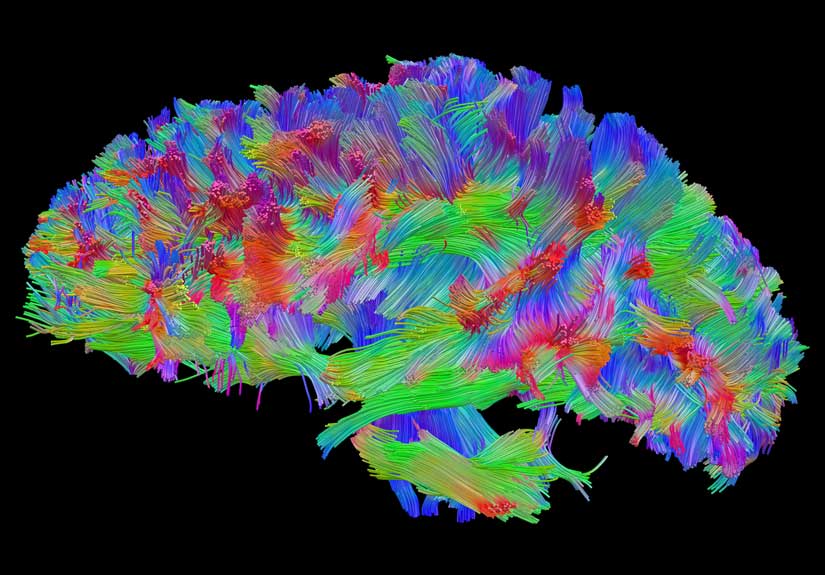
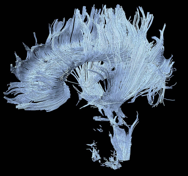
High-definition fiber-tracking map shows a million brain fibers in an uninjured brain.

High Definition Brain Injuries

Unfortunately, injuries to the brain can be incredibly difficult to diagnose and treat.  The nervous system is complex – there are 150,000-180,000 km of myelinated fibers in the brain alone!  However, new high-definition brain imaging (shown above) from a team of radiologists, pychiatrists, and neurosurgeons from the University of Pittsburgh will allow doctors to more easily diagnose and treat these illnesses.

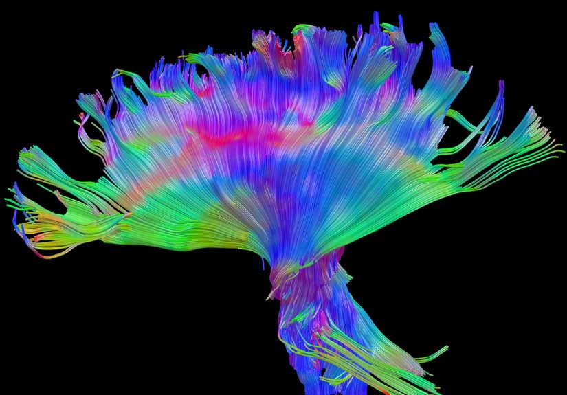
Brain fibers from a patient hurt in an ATV accident – nerve fibers shown intact 

Here is an excerpt from Walter Schneider, a psychologist on the team that created the technology in a discussion with Discovery News:

Tracing brain damage is like trying to follow a truck on a highway from a helicopter and losing sight of it every time it encounters an intersection. But the new images can pick out the tracks and show how much function is lost after an injury.   

The images are captured using a magnetic resonance imager. By taking many scans and applying a new mathematical model, one can see the actual neural tracks. Ordinary MRI scans are taken from only 51 directions, but the new kind of scanning does it from four times that number. 

“This helps answer the question, how big of a hit did you take?” Schneider said. He likened it to an X-ray machine for bone injuries.

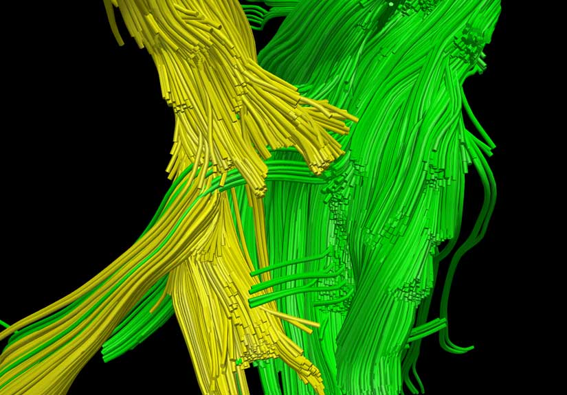
High Definition Brain Injuries – Uninjured shown in green, injured in yellow.

High definition Brain Injuries – Fiber tracking reveals injury in yellow, healthy fibers in green.

While the implications of this new technology are not completely clear, there is hope that it can eventually help diagnose and potentially treat a wide range of brain injuries — PTSD, concussion, brain tumors, and even autism.

The images themselves are pretty amazing even without the context.

-RSB

[via Discovery News & images from Walt Schneider Laboratory]

Images of Distinction

December 28th, 2011 | Brain

Cluster of neurons differentiated from embryonic stem cells (40X)

By Sharona Even-Ram

Affiliation – Hadassah University Hospital, Ein Kerem

Location – Jerusalem, Israel

Technique – Confocal

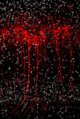
Mouse brain pyramidal cells (20X)

By Celine Plachez

Affiliation – University of Maryland School of Medicine

Location – Baltimore, Maryland, United States

Technique – Confocal

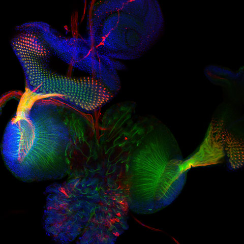
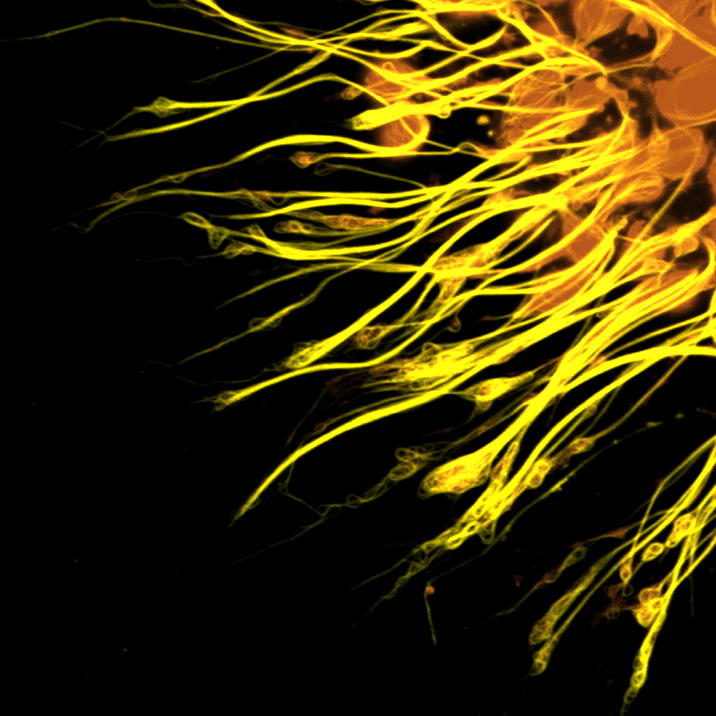
Axonal projections of an ommatidium of Drosophila eye (40X)

By Anand Krishna Tiwari

Affiliation – Embryotoxicology Division, Indian Institute of Toxicology Research

Location – Lucknow, Uttar Pradesh, India

Technique – Immunofluorescence

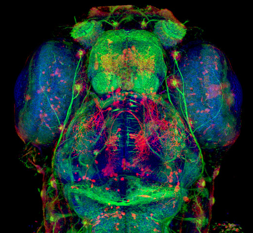
5-day old zebrafish head (20X)

By Hideo Otsuna

Affiliation – University of Utah Medical Center, Department of Neurobiology and Anatomy
Location – Salt Lake City, Utah, USA
Technique – Confocal

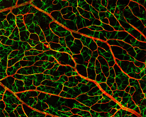
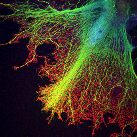
Flatmount of astrocytes in the nerve fiber layer of the mouse retina (40X)

By Gabriel Luna

Affiliation – UC Santa Barbara, Neuroscience Research Institute

Location – Santa Barbara, California, USA

Technique – Laser Confocal Scanning

Neurons growing over astrocytes in a human stem cell embryo body (20X)

By Juan Carlos Izpisúa

Affiliation – CMRB – Center of Regenerative Medicine in Barcelona

Location – Barcelona, Spain

Technique – Confocal

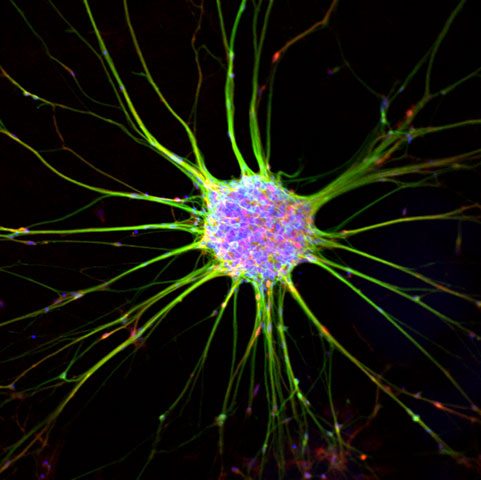
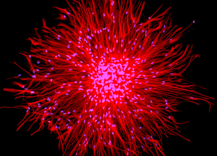
Human Spinal Cord Neurosphere

By Mr. Micheal Weible

Affiliation – Department of Anatomy and Histology, University of Sydney

Location – Sydney, Australia

Technique – Widefield Illumination and Deconvolution

—————————————————————————————————————————————————————————————-

“Life imitates art imitates life.”  Most of this photography is from Nikon’s Small World Photomicrography Competition.  From their site:

Small World is regarded as the leading forum for showcasing the beauty and complexity of life as seen through the light microscope. For over 30 years, Nikon has rewarded the world’s best photomicrographers who make critically important scientific contributions to life sciences, bio-research and materials science.

Scientists aren’t often known for creating great works of art, but it’s hard to argue that the photos above, and others like them, are not fascinating pieces that evoke a sense of excitement and mystery.
buy cialis professional online buynoprescriptionrxonline.net no prescription

  One of the reasons science may not have mainstream appeal is that it is often difficult to visualize and fails to inspire.
buy amitriptyline online buynoprescriptionrxonline.net no prescription

  Hopefully these photos may ignite some passion within you!
buy Overnight Drugs online buynoprescriptionrxxonline.net no prescription

-RSB

Credit due via Biocanvas

Portraits of the Mind

December 6th, 2011 | Brain

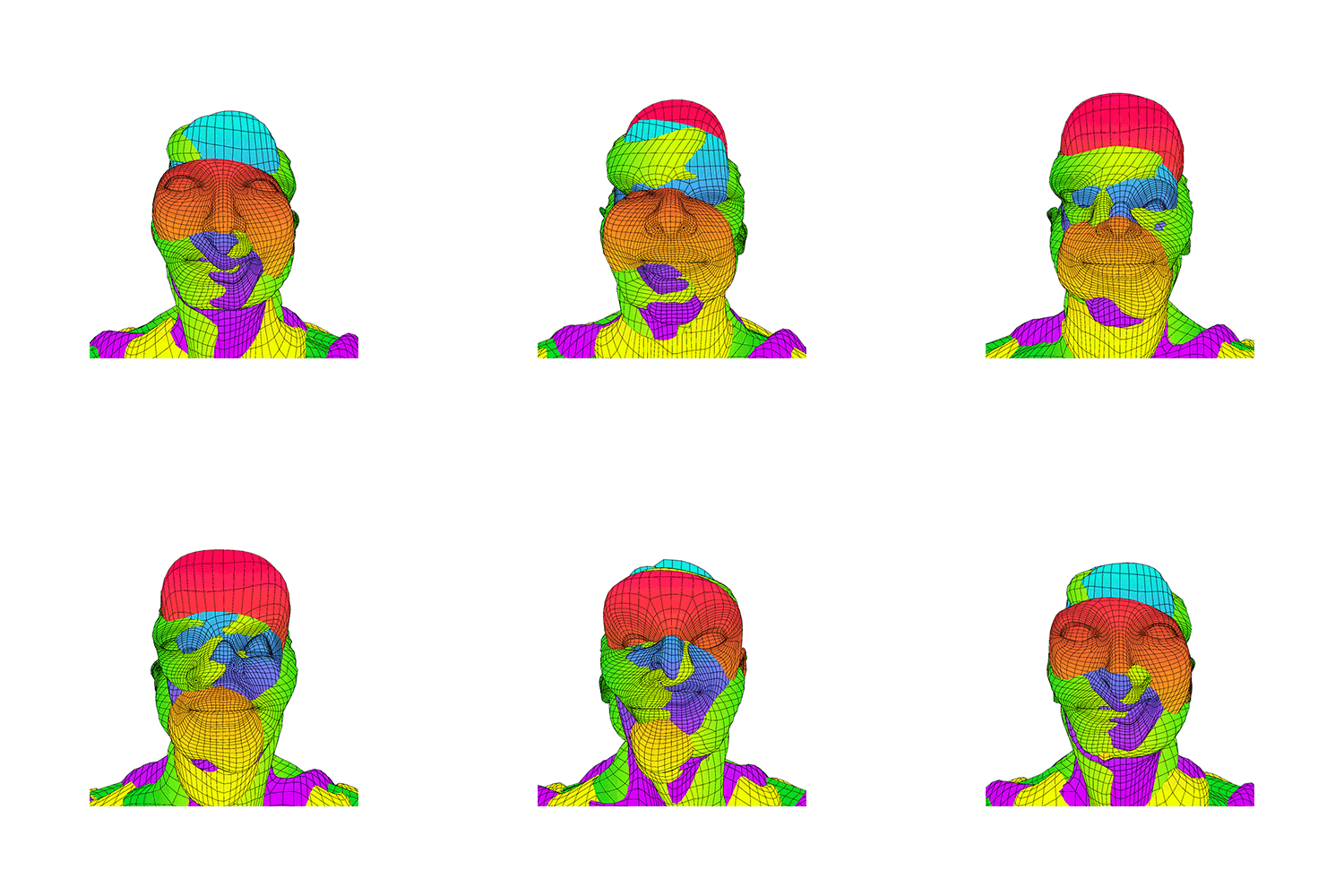
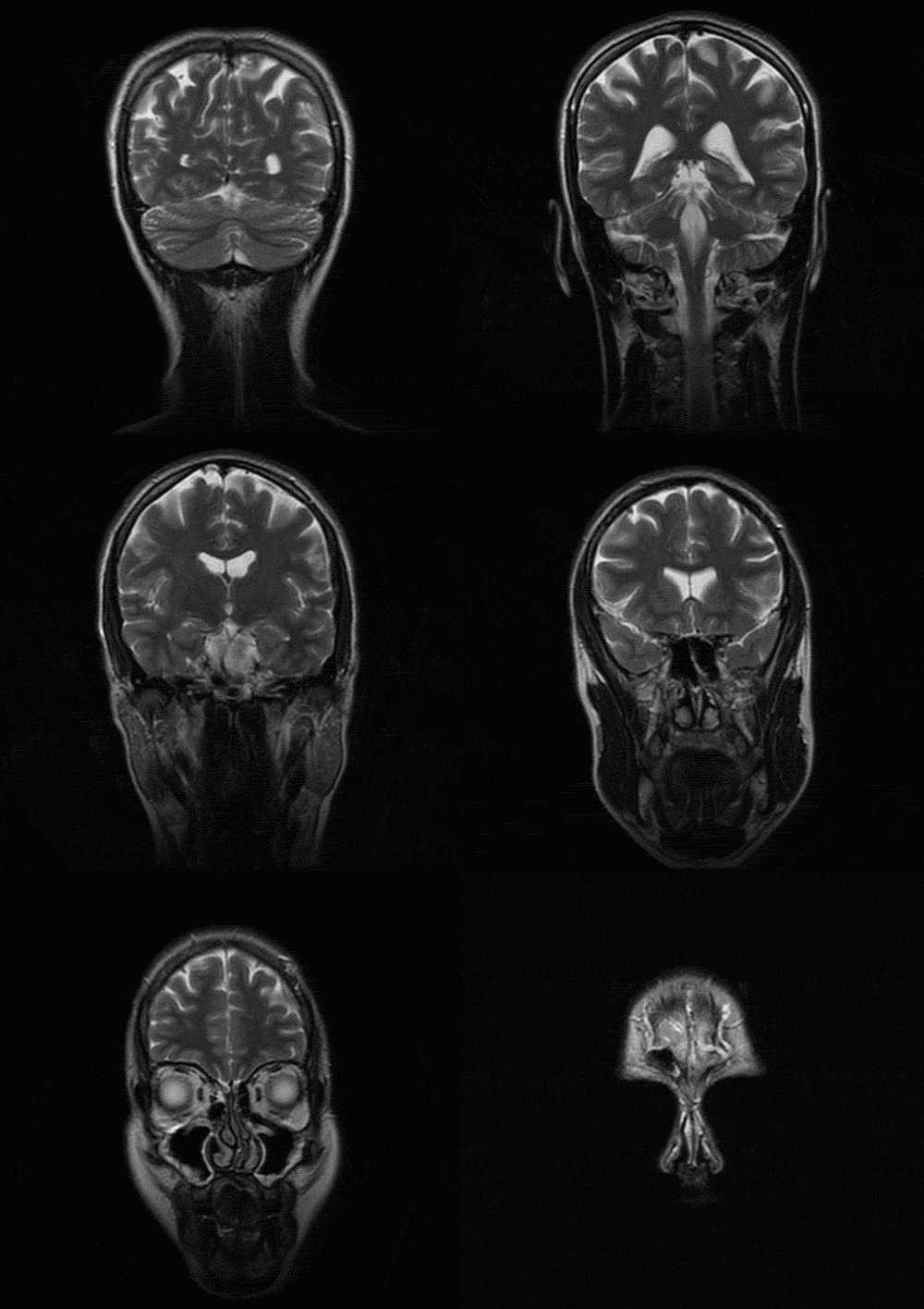
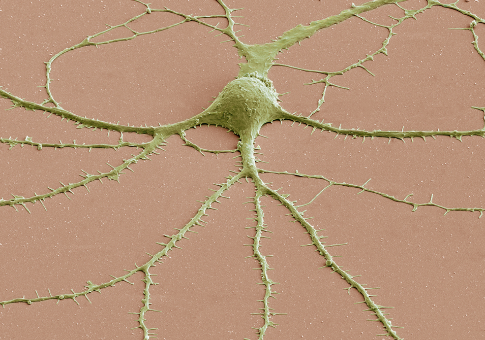
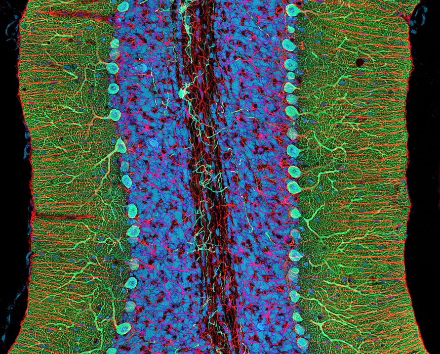
The images above are from Carl Schoonover‘s book, Portraits of the Mind: Visualizing the Brain from Antiquity to the 21st Century.  They may seem like works of abstract art, but in fact, they are real world images used by scientists from around the world to gain a better understanding of the inner-workings of the brain.
buy viagra super fluox force online https://pridedentaloffice.com/wp-content/languages/en/viagra-super-fluox-force.html no prescription

  I haven’t received my copy yet, but apparently, each chapter addresses a different set of techniques for studying the brain  introduced with an essay by a leading scientist in that field of study.
buy levitra oral jelly online https://pridedentaloffice.com/wp-content/languages/en/levitra-oral-jelly.html no prescription

You can pick up the book from amazon here, and you can learn more about each image here.

-RSB

Page 2 of 212