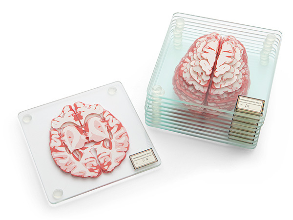
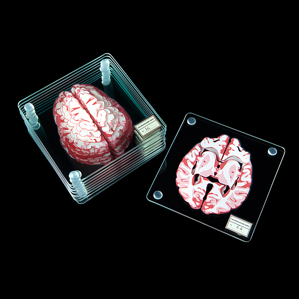
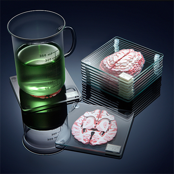
If you want to nerd out your house guests, look no further. The folks at Think Geek put together this simple, yet cerebral idea :). buy prelone generic gaetzpharmacy.com no prescription
Each coaster resembles an axial slice of a human brain. In other words, if you stack them all together in the right order, you get a recreation of the brain.
I’m having neuroanatomy lab flashbacks. buy lasix generic gaetzpharmacy.com no prescription
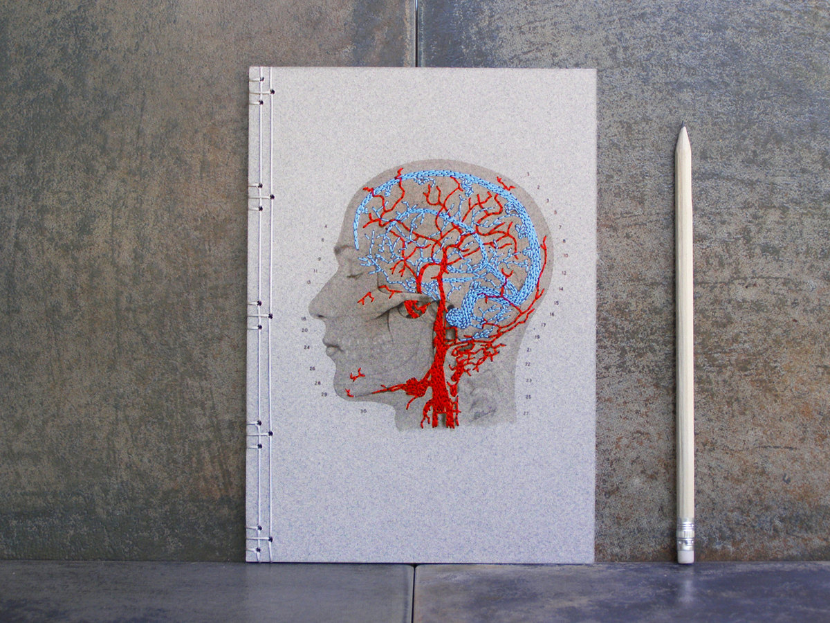
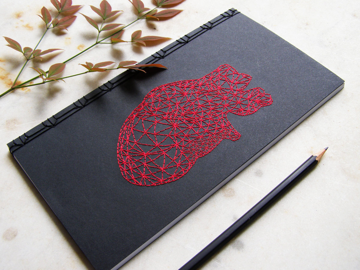
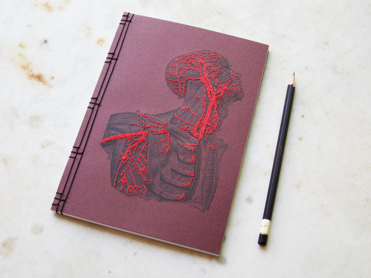
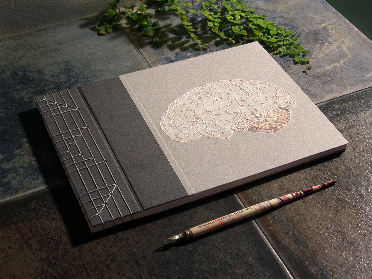
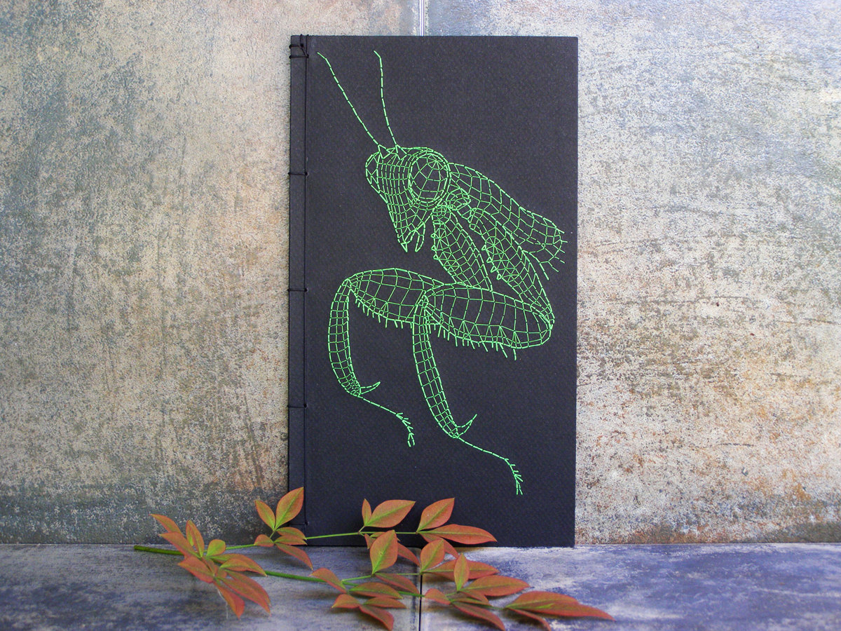
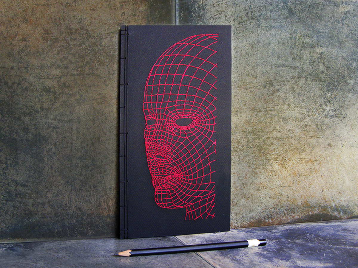
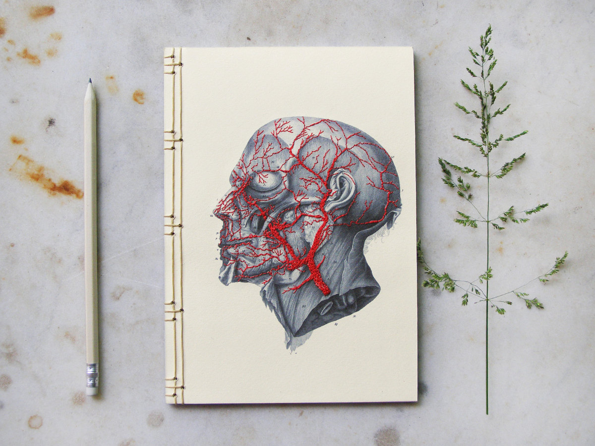
The folks at Fabulous Cat Papers in Athens, Georgia have put together an incredible collection of hand embroidered notebooks featuring a bunch of beautiful designs. The classic anatomical illustrations really come to life with the addition of red and blue (arteries and veins, respectively) thread accents.
Welcome to “A Journey Through The Visual System”! I made this video to promote brain awareness for the general public. As a neuroscience researcher, I’ve always believed it’s important to help people understand the complexities of the human nervous system. Thus, I hope this video can be appreciated by kids and adults alike, and something will be learned by all.
The project was inspired by the old Magic School Bus show I used to love as a kid. In the video, you will go on a 5-minute journey from the eye all the way to brain, learning the neuroanatomy along the way.
If you are feeling kind, you can vote for the video here (before Sept. 30th). This was submitted as part of a contest hosted by the Society for Neuroscience and BrainFacts.org.
Anyway, thanks for watching!
Here is the script for the video in case you missed something important:
“We begin our journey with the eye, specifically the iris, which gives the eyes its distinctive color. Now, the iris can be green, or blue, or brown, or black depending on the level of melanin which it contains. When the lights go off, the muscles connected to the iris contract, which makes the dark circle in the center of the eye, the pupil, get bigger. When the light goes on, the pupil gets smaller to allow less light to enter. This is how the eye adapts to light.
Ahh, that must be Ellie. She’ll be our tour guide on this journey through the visual system. Hi Ellie, how are you? To get a better look at the visual system, we’ll need some light. Let’s observe the anatomy of the eye in a little bit more detail. If we peel away the skin, we can see the arteries and veins, which supply important nutrients to the area. Next, we can see the surrounding muscles, which help move the eye in all directions. Now, let’s cut the eye in half to see how light enters the visual system. First, it hits the cornea, the protective layer of eye. Then, through the whole in the iris known as the pupil. Lastly, the job of the lens is to bend light to focus it correctly on the retina.
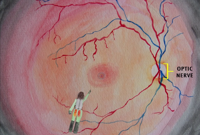
It’s time to enter the eye. Ellie has her jetpack and now we’re looking at the surface of the inside of the eye. The optic nerve on the right is where all the nerve fibers leave the eye heading toward the brain. Let’s follow Ellie has she heads toward that dark spot in the distance. This is the fovea centralis, a small pit in the retina responsible for our sharpest, clearest vision. Foveal vision allows us to do things like read books, or drive cars, or play video games.
Now, let’s look at a cross-section of the retina to see how neurons respond to light. Light is absorbed by rods and cones, which are specialized photoreceptors. This starts a chain reaction, which excites the bipolar cells and then subsequently the ganglion cells, to send electrical signals off toward the brain. The amacrine and horizontal cells work to modulate the circuit. buy cialis sublingual online https://healthcoachmichelle.com/wp-content/languages/en/cialis-sublingual.html no prescription
Visual information flows along the optic nerve like a river of electricity. At the optic chiasm, the signals split such that images from the left visual field head to the right brain, and images from the right visual field head to the left brain.
After the optic chiasm, the visual signals make a quick stop at the lateral geniculate nucleus, or LGN. The LGN is organized into 6 layers, which all receive extensive feedback control from higher visual areas.
From the LGN, the visual signals travel along optic radiations back to the visual cortex. The cortex is where we use the signal that originally came from the eye to construct our visual reality. The billions of neurons in the human brain work to encode and process the information. Information is sent forwards and backwards. See, the beauty of the visual system is that everything we see is affected by our memories, and our feelings, and what we’ve seen before.
Well, that concludes our journey through the visual system, see you next time.”
The scientific paper they are all referring to is linked here (although you can’t read it without a subscription, which brings up a totally different problem in access to science):
When I first read these headlines, I was understandably intrigued. Had someone REALLY found good evidence in MRI data that there are differences in the brains of casual marijuana smokers? The idea is not totally far fetched. Alcohol is known to be neurotoxic and it can shrink the size of the brain through dehydration, but this is mostly corrected after you quit drinking. Carbon monoxide in cigarette smoke is also a known neurotoxin, but I’ve never read about any changes in the size of anatomical regions of the brain from smoking tobacco, unless you count brain tumors. However, no one has ever really found evidence to support major anatomical changes in the brain following marijuana use. So could this really be true?
Even before reading the paper, my intuition said the answer was no… brain imaging research is notoriously fraught with spurious findings linked to inappropriate use of statistics.
I gave the paper a casual read, and immediately, I noticed problems, MAJOR problems. First of all, sample size… only 20 people were included in the cross-sectional study. That is low. A cross-sectional study means that they had no within-subject comparisons. In other words, all of the data was collected at one time. A much stronger approach would have been to image subjects before they smoke and then through time as they begin to “casually smoke” marijuana. Of course, this is much more difficult, but with a study design containing so many potential confounds (see below), it’s pretty much required (imho) to say anything definitively.
Second of all, the confounds… the investigators did not control for various other aspects of these people’s lives that may cause changes in brain anatomy. How much did each subject drink? smoke tobacco? do other drugs? etc… These all could be equally correlated to the differences in brain anatomy which they discovered. Or it could something entirely different like genetics?
And lastly, the statistics… I came across this article by computational biologist Lior Pachter, and that was sort of the nail in the coffin. I suggest reading through it because Lior does a great job of highlighting the problems with multiple comparison statistics, causation vs. correlation, and many other mistakes.
He even calls it “quite possibly the worst paper [he’s] read all year.” The Journal of Neuroscience is a rather prestigious journal, so this is all the more upsetting.
I do not study the effects of marijuana use on the brain, so I can’t tell you how it may or may not cause harm. I am absolutely positive it has some effect. But it’s important to remember that pretty much everything you do creates changes in your brain. Reading a book, riding a bike, talking with your friends… buy kamagra soft online https://www.mabvi.org/wp-content/languages/en/kamagra-soft.html no prescription
these all create lasting memories that are encoded in your neurons. However, after reading this most recent article, there is ABSOLUTELY NO WAY you can assert that marijuana use is harmful, or creates anatomical changes, or anything really…