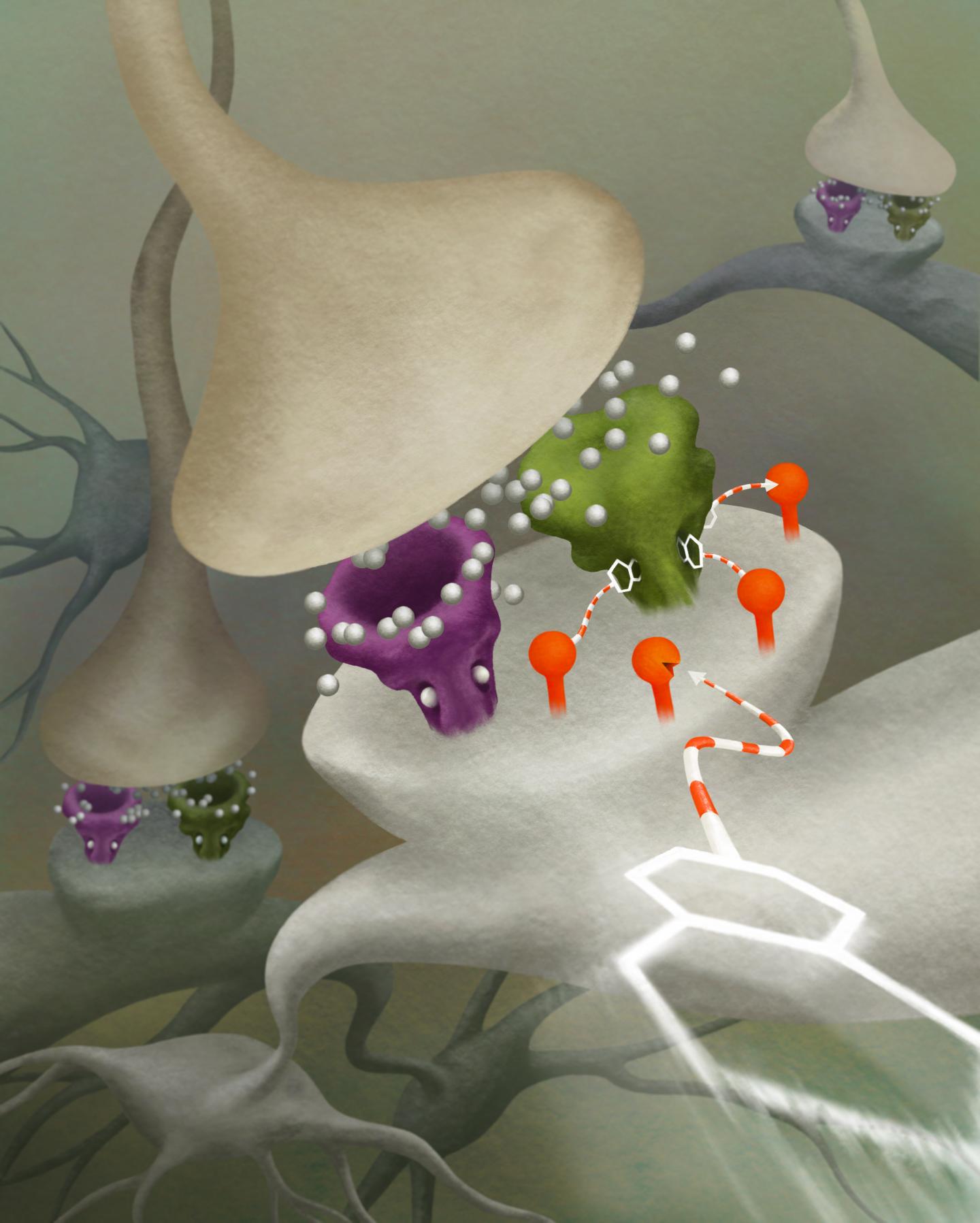
Julia Kuhl is an artist who has worked for a variety of independent science labs, top-tier academic journals, as well as a host of news organizations (The New Yorker, Chicago Magazine, et cetera). She has illustrated and animated a diverse set of topics ranging from neural populations to viscous flow states. buy super avana online https://www.conci.com/wp-content/languages/en/super-avana.html no prescription
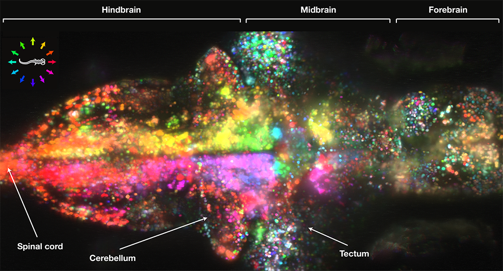
A team led by Drs. Jeremy Freeman and Misha Ahrens recently recorded the activity of approximately 80,000 neurons firing in the brain of a zebrafish larvae. The technique they implemented is called light-sheet microscopy. Briefly, the scientists genetically engineer zebrafish neurons to emit a fluorescent signal just after the neuron fires. Laser beams are the shot through the fish so that the activated neurons will glow and an overhead microscope records the whole thing. Of course, this technique only works because the zebrafish are entirely transparent, so don’t expect to have your brain scanned in this manner any time soon.
“At the beginning of the movie, the fish is resting and the forebrain region on the far-right is flashing away. That may represent whatever the fish is thinking about when it’s just hanging out.
Its intent to swim to catch up was measured with electrodes on its muscles. When the bars start sliding, a few neurons sitting just behind the eyes light up followed by a huge cascade of activity, including massive pulses initiating swimming.”
“There must be fundamental principles about how large populations of neurons represent information and guide behavior,” says neuroscientist Jeremy Freeman of Janelia Farm Research Campus in Ashburn, Virginia. “In this system, where we record from the whole brain, we might start to understand what those rules are.”
We know that the processing of sensory input and the generation of behavior involves large networks of neurons, and Dr. Freeman believes that observing networks with this sort of technology will enable us to gain deeper insight to how the brain functions. buy grifulvin online https://www.conci.com/wp-content/languages/en/grifulvin.html no prescription
It is important to note that the temporal resolution is fast enough to identify which neurons are involved in a given behavior but too slow to count how many times they fire. Thus, there is no way that this technique could ever decipher the neural computations that take place at the millisecond timescale in the human brain.
Small World is regarded as the leading forum for showcasing the beauty and complexity of life as seen through the light microscope. For over 30 years, Nikon has rewarded the world’s best photomicrographers who make critically important scientific contributions to life sciences, bio-research and materials science.
Scientists aren’t often known for creating great works of art, but it’s hard to argue that the photos above, and others like them, are not fascinating pieces that evoke a sense of excitement and mystery. buy cialis professional online buynoprescriptionrxonline.net no prescription
One of the reasons science may not have mainstream appeal is that it is often difficult to visualize and fails to inspire. buy amitriptyline online buynoprescriptionrxonline.net no prescription
Hopefully these photos may ignite some passion within you! buy Overnight Drugs online buynoprescriptionrxxonline.net no prescription
The images above are from Carl Schoonover‘s book, Portraits of the Mind: Visualizing the Brain from Antiquity to the 21st Century. They may seem like works of abstract art, but in fact, they are real world images used by scientists from around the world to gain a better understanding of the inner-workings of the brain. buy viagra super fluox force online https://pridedentaloffice.com/wp-content/languages/en/viagra-super-fluox-force.html no prescription
I haven’t received my copy yet, but apparently, each chapter addresses a different set of techniques for studying the brain introduced with an essay by a leading scientist in that field of study. buy levitra oral jelly online https://pridedentaloffice.com/wp-content/languages/en/levitra-oral-jelly.html no prescription
You can pick up the book from amazon here, and you can learn more about each image here.
Greg Dunn is a 6th year Neuroscience PhD student at the University of Pennsylvania and he is responsible for these wonderful paintings inspired by minimalist scroll and screen painting from the Edo Period in Japan. He paints neurons from different parts of the nervous system in the Asian sumi-e stytle and the results are amazing. He’s been commissioned to do pieces for a handful of Neuroscience departments around the country. buy intagra online https://www.calmandgentledentalcare.co.uk/wp-content/languages/en/intagra.html no prescription


")
")
")
")
")


