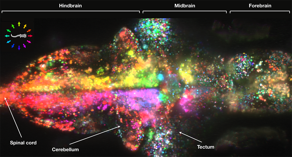
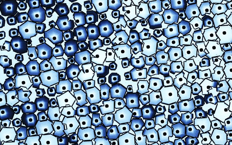
“Nanoscale Lego Puzzle” – Radha Boya
The hidden beauty of the natural world is brought forth in laboratories around the country on a daily basis. Unfortunately, the general public usually doesn’t get the chance to see these incredible scientific images. However, Northwestern University has recently been holding a photo competition to share images “across a wide range of disciplines, including medicine, chemistry, engineering and nanotechnology.” They are truly a sight to behold.
buy sildenafil tadalafil vardenafil online https://www.mydentalplace.com/wp-content/languages/en/sildenafil-tadalafil-vardenafil.html no prescription
Seen above is an image called “Nanoscale Lego Puzzle” by Radha Boya. Here’s a description of the piece:
“A thin film of gold has been deposited on a silicon mold. Each evenly spaced black dot is a groove in the silicon that has been filled by the gold.
buy free viagra online https://www.mydentalplace.com/wp-content/languages/en/free-viagra.html no prescription
The puzzle-like shapes are made when the gold cracks and curls up. The darker shading indicates where the gold has curled in on itself. The metal tips fabricated by this method can be used in many applications, such as printing DNA chips.”

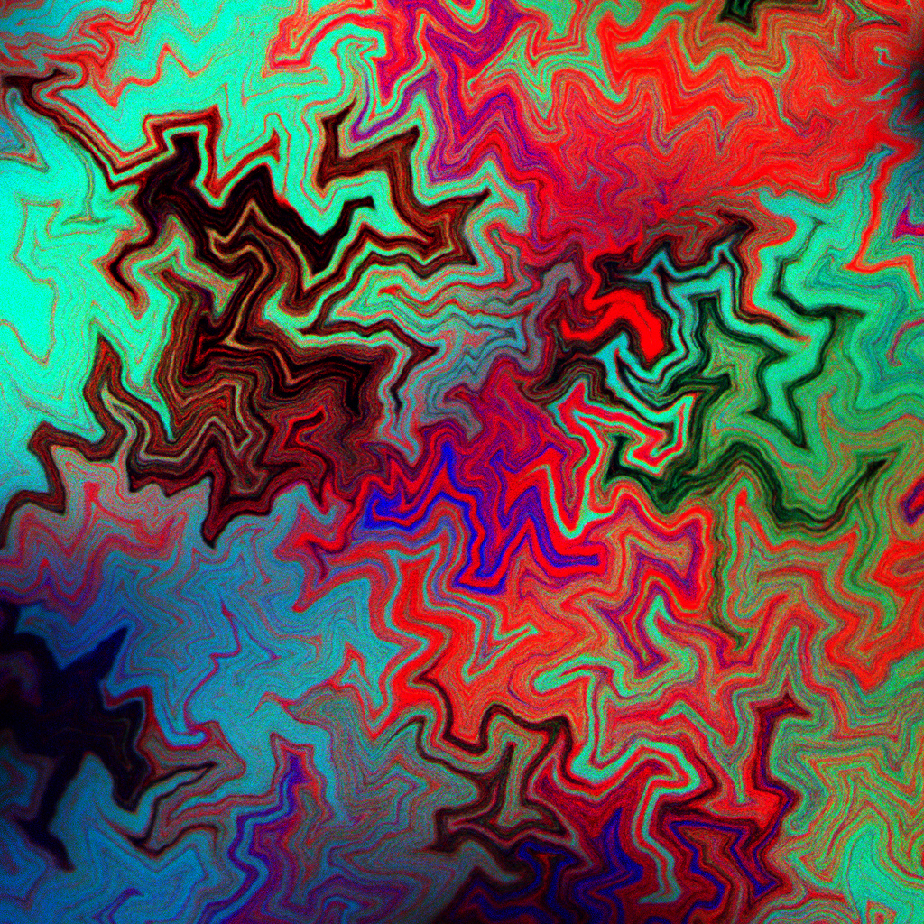
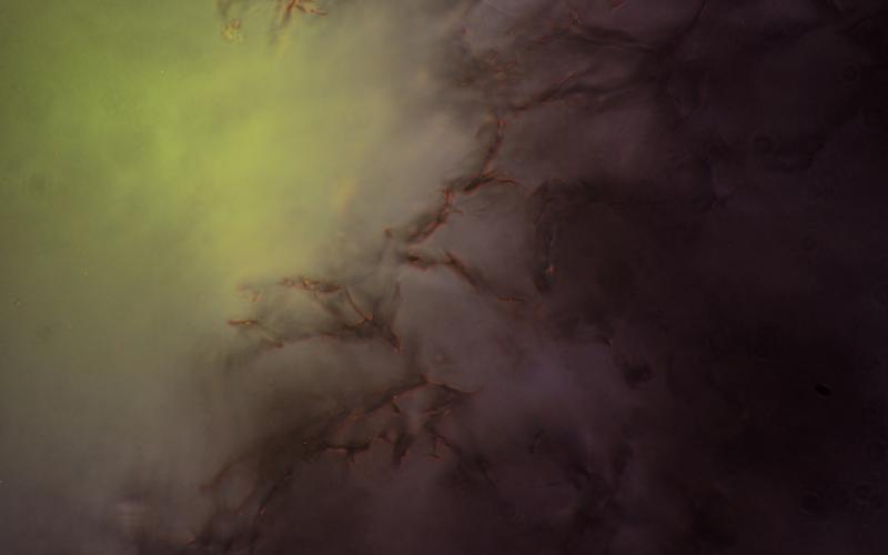
“Meltdown” – Keith Brown
“This image shows the aftermath of a case when a large voltage was applied between a thin film of gold (light blue) and silicon (dark blue), and electrons found a way through the thin insulating layer that separated them. As the current increased, the material was heated resulting in a catastrophic thermal expansion that resulted in a crater of solidified material (red).”
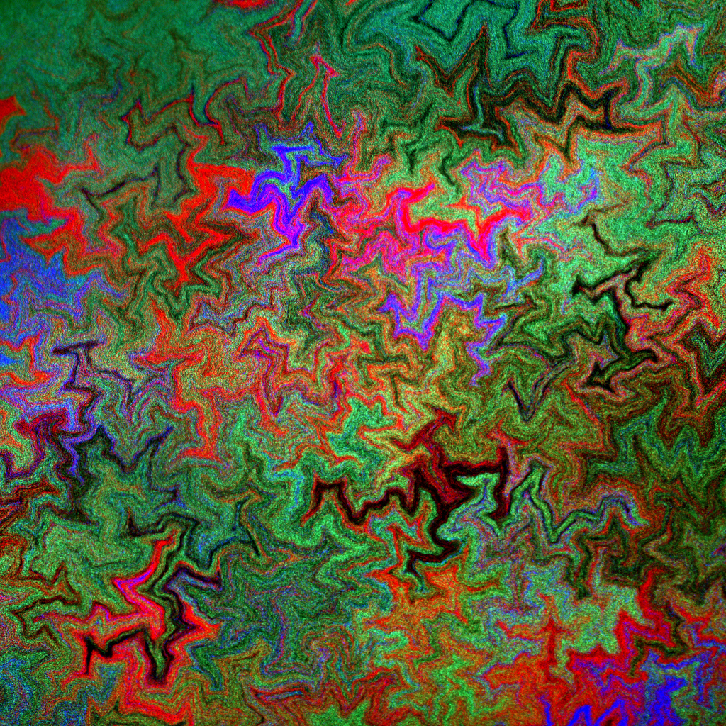
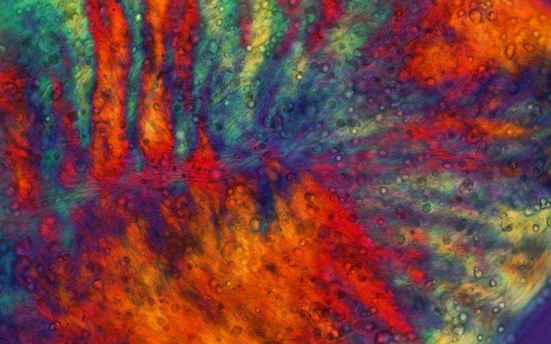
“Graphene Oxide” – Andrew Koltonow
“Koltonow and his colleagues study a material called graphene oxide (GO), which is only a couple of atoms thick. They can assemble thin sheets of GO into a foam that conducts electricity. The foam can be used to create electrodes for batteries, making such energy storage devices smaller and lighter.
In this image, graphene oxide sheets (purple-orange) cast shadows from light that is scattered off of GO foam (green-yellow), creating an eerie effect.”
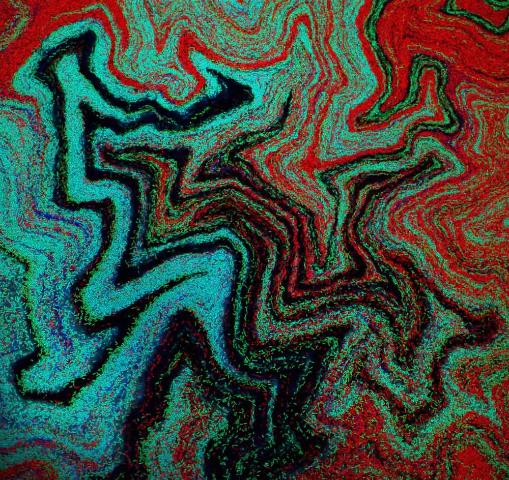
“Colorful Directions” – Mark McClendon
“Imagine simply injecting healthy human cells into the body to repair damaged muscle tissue. This might one day be possible if scientists, like McClendon, can find a way to keep these cells organized at the point of injury until healing is complete. One solution might be placing the cells in a nanofiber gel, which can then be injected directly into the human body. The cells growing in the gel will eventually respond to their surroundings by stretching and migrating in the same direction as the nanofibers.
Shown in this image is a blob of nanofiber gel with encapsulated cells from a human heart. The color is a result of the alignment of nanofibers making up the gel, with each color corresponding to a cluster of nanofibers aligned one direction.”
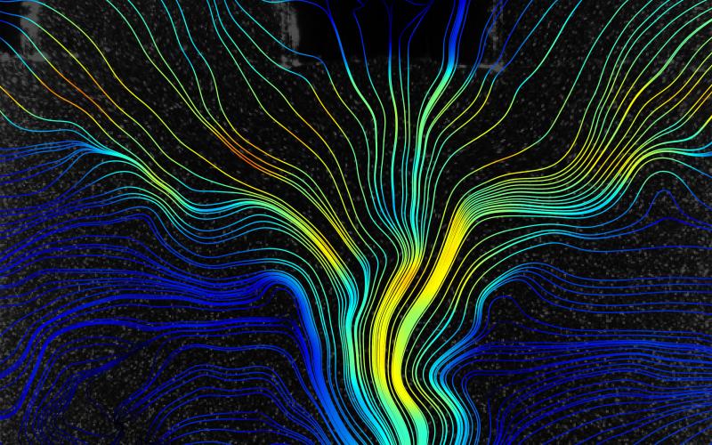
“Black Ghost Knifefish” – Oscar Curet
“The black ghost knifefish, found in the Amazon Basin, can move rapidly and in many directions due to an elongated fin on its belly that runs nearly the entire length of the fish. Curet and his colleagues used a robotic replica of the knifefish to study this motion, which could prove useful when applied to the design of underwater vehicles like submarines.
This image maps the motion of the robotic fish as it moves in a vertical direction. The lines represent the path of the fluid motion and the color represents the velocity, where blue is slow and yellow is fast.
buy augmentin online https://www.mydentalplace.com/wp-content/languages/en/augmentin.html no prescription
“
The mysteries of science can be illuminated with a well-captured photo or illustration. I’m glad Northwestern is sharing some of this wonder with the general public.
You can check out the full list of winning scientific images at the Northwestern website.
-RSB