A new exhibition on the history of anatomy, Body of Knowledge, opened recently at Harvard and will be on display until December, 2014.
From the Harvard Museum of Science & Culture:
“Body of Knowledge” will explore the act of anatomizing not as a process of mapping a finite arrangement of bodily structures, but as a complex social and cultural activity. By means of a diachronic perspective, the exhibit narrative cuts through the multiplicity of anatomical practices, presenting three important moments in the history of anatomy: sixteenth century dissections and anatomical drawings, nineteenth century anatomical practices, and contemporary use of both cadavers and digital technology for anatomic education. “Body of Knowledge” hopes to capture the complexity of the many people, places, and meanings involved in human dissection.
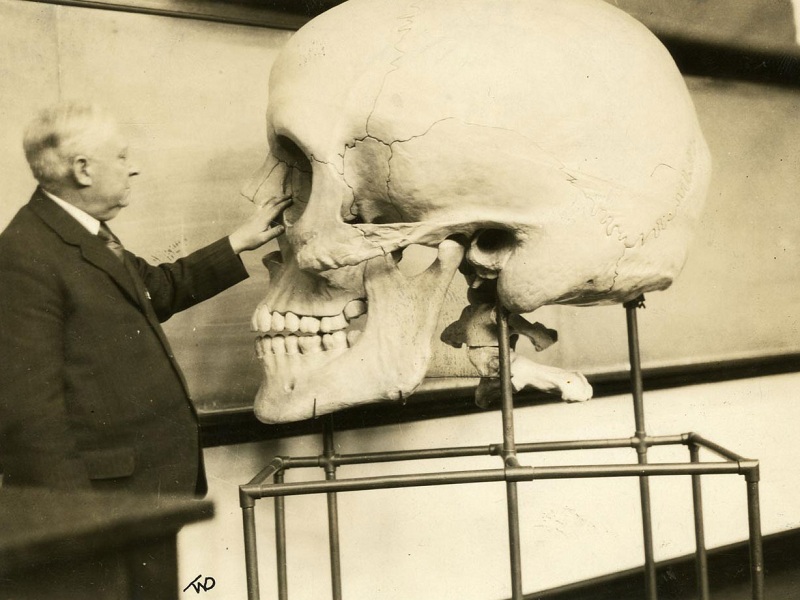
Seen above is Harris P. Mosher lecturing at Harvard Medical School in 1929. The giant skull was made in the 1890s and is a piece in the new exhibit. I’d love to have that on display in my living room!
While perusing the archives of one of my favorites inspiration sources, Brain Pickings, I came across these century-old anatomy illustrations made by E.J. Stanley. The images cycle through three main layers of the human body — skin, muscle and bone. I’ve always appreciated the style of illustration used in old anatomy texts, and a flip book is a great way to demonstrate the subject. The illustrations really remind me of the old French anatomy plates created by Gautier D’Agoty.
If you enjoyed these, you might be able to pick up some old posters by E.J. Stanley on EBay Here.
This incredible pristine brain specimen is fresh out of Anatomy class at the University of Utah. You can even hear the bone saws humming along in the background! The brain has just been removed from an autopsy of a cancer patient who donated his/her body to science.
As Dr. Suzanne Stensaas points out in the video, the take home message is that human brain is extremely soft and squishy. So wear your helmet!
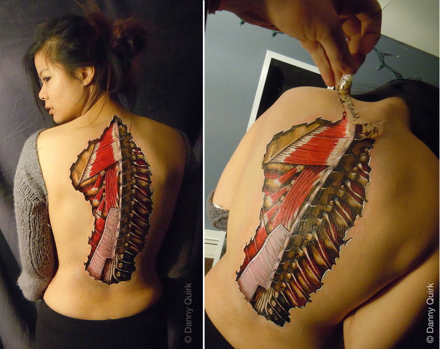
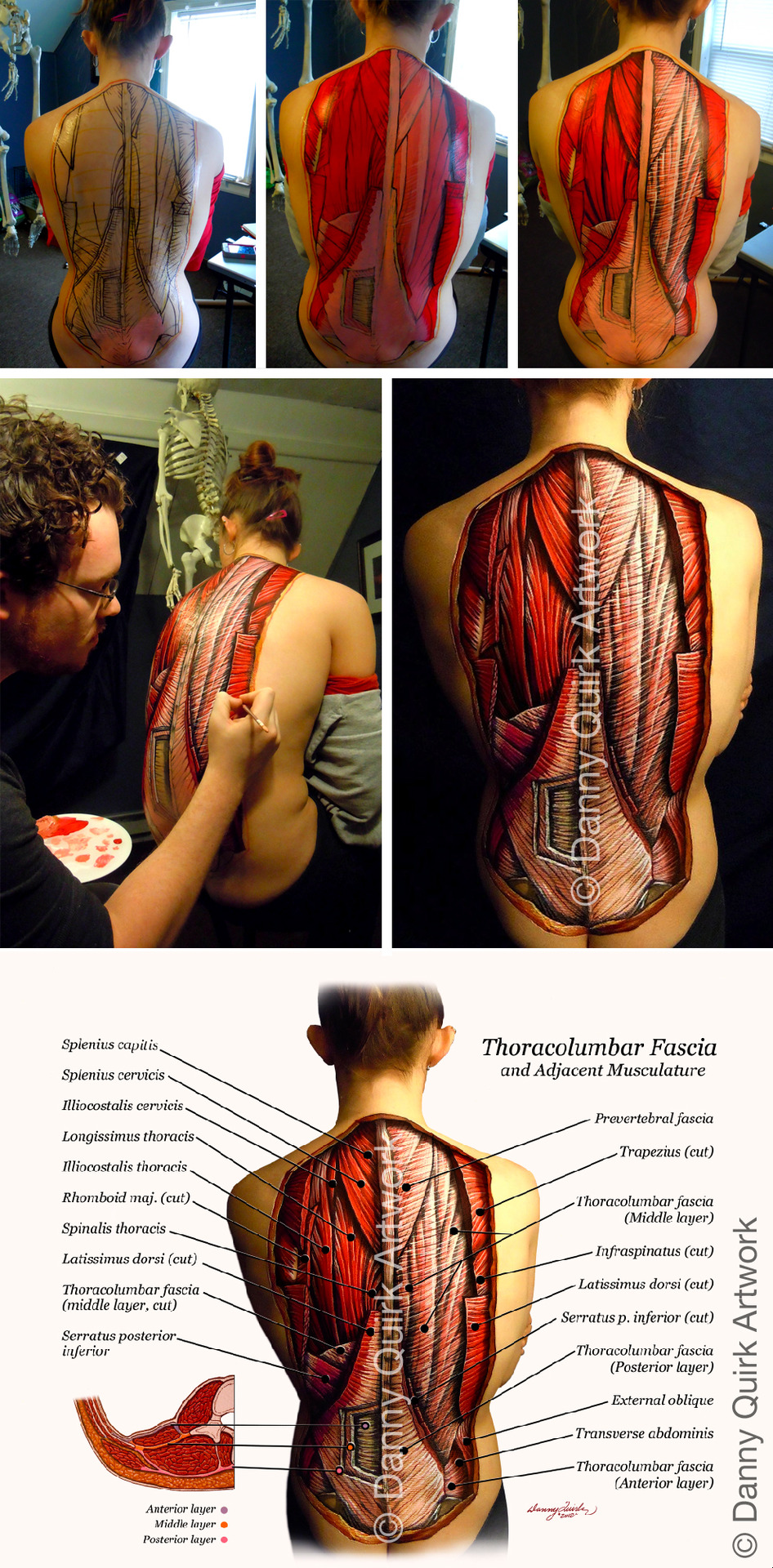
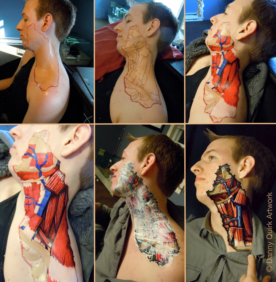
Danny Quirk is a rising star in the field of anatomical art. He created these beautiful and educational body paintings, which take roughly 6 hours and are composed of latex, sharpie pen, and acrylic paint. Apparently, he stumbled upon the concept while trying to make his girlfriend a Halloween costume… buy levitra soft online https://blackmenheal.org/wp-content/languages/en/levitra-soft.html no prescription
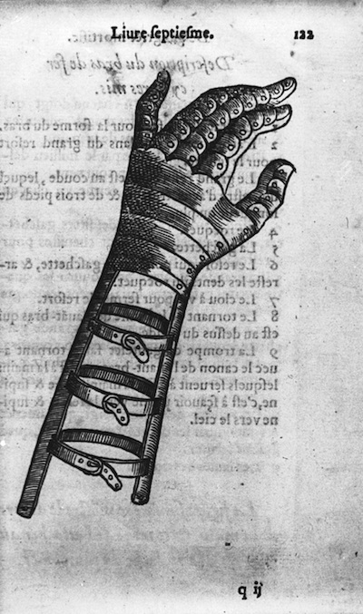
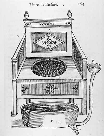
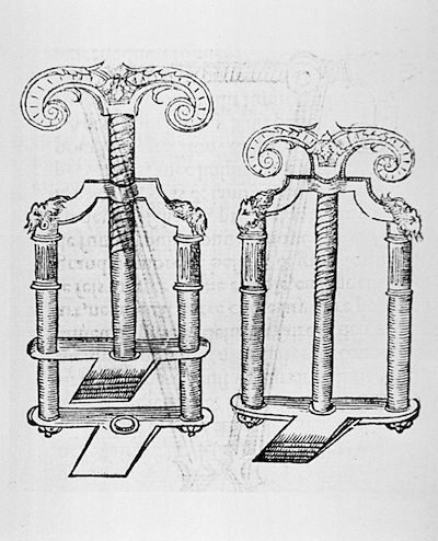
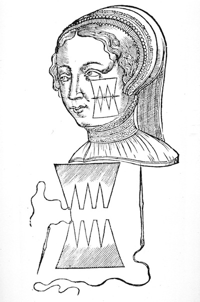
And here are a few other illustrations from Dix livres de la chirurgie:
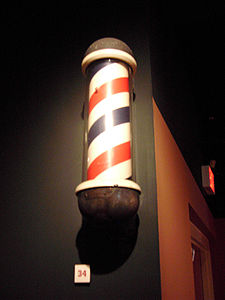
The images above are mechanical prosthetics as designed by Ambroise Paré in his book Dix livres de la chirurgie (Ten books of Surgery). Paré was a French barber surgeon and is considered to be one of the fathers of surgery and modern forensic pathology. Interesting fact: the barber pole is a vestige of an era when barbers were the primary surgeons and NOT physicians. buy viagra professional online https://www.calmandgentledentalcare.co.uk/wp-content/languages/en/viagra-professional.html no prescription
Paré was the official royal surgeon for kings Henry II, Francis II, Charles IX and Henry III. He designed a range of surgical instruments and was a leader in surgical techniques and battlefield medicine, especially the treatment of wounds. He amputated countless limbs during his career as thousands of French cavalrymen were being killed and wounded by enemy arquebuses. The prosthetics above appear quite advanced for the 16th century. The hand has complex finger movements which modern prosthetics often fail to include. buy fildena-xxx online https://www.calmandgentledentalcare.co.uk/wp-content/languages/en/fildena-xxx.html no prescription
He also made original artificial eyes from enameled gold, silver, porcelain and glass.
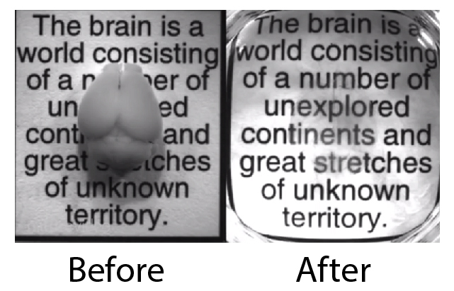
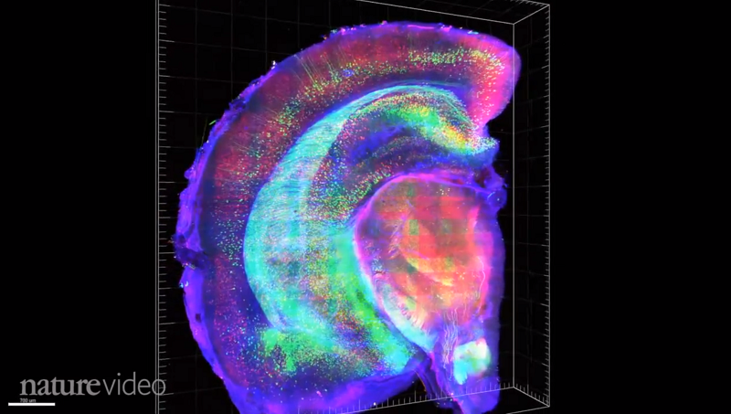
The world’s very first See-Through Brain has been developed by a team at Stanford University led by Karl Deisseroth (M.D., Ph.D.). Deisseroth is well-known for his critical role in the development of Optogenetics, a tool used to control individual neurons with light. Optogenetics is normally limited to surface neurons because the light has trouble reaching deeper areas, but the see-through brain may greatly enhance its efficacy.
The new method (termed CLARITY) involves removing the fat that provides structure but also blocks light. The brain is soaked in a chemical that forms a nanoporous hydrogel-hybridized mesh in the brain. This mesh can then support all the tissue so the fat can be washed away, resulting in the incredible see-through brain.
Unfortunately, the new technique can’t be used in living animals, but it still represents a huge advancement for neuroanatomists. No longer will there be much need to cut the brain into tiny slices (an extremely time-consuming process) to observe connectivity.
The announcement comes just a week after President Barack Obama announced a $100 million BRAIN initiative, and this new step forward surely offers a taste of the sort of technological breakthroughs the initiative hopes to achieve.
And all the Leaders in Neuroscience seem to be weighing in on this one:
“I can’t make any official statement, but I can say that this is exactly the type of technology one would hope to develop for the [BRAIN] project” – Dr. Michelle Freund, a program manager with the National Institutes of Mental Health
“If the entire mouse brain is transparent, that makes a very large fraction of neuroscience research much easier” – Dr. R. Clay Reid of the Allen Institute for Brain Science in Seattle.
This technique “is a giant step forward from having to slice the mouse brain into 1,000 pieces and looking at them each individually, then trying to reconstruct the relationships of all those slices” – Dr. Cori Bargmann of Rockefeller University, a co-leader of Obama’s brain initiative.
“It’s exactly the technique everyone’s been waiting for”- Dr. Terry Sejnowski of the Salk Institute.
Karl Deisseroth, mastermind of the CLARITY technique
It is certainly an exciting time to be a Neuroscientist.